Magnetic resonance imaging (MRI) of the spine
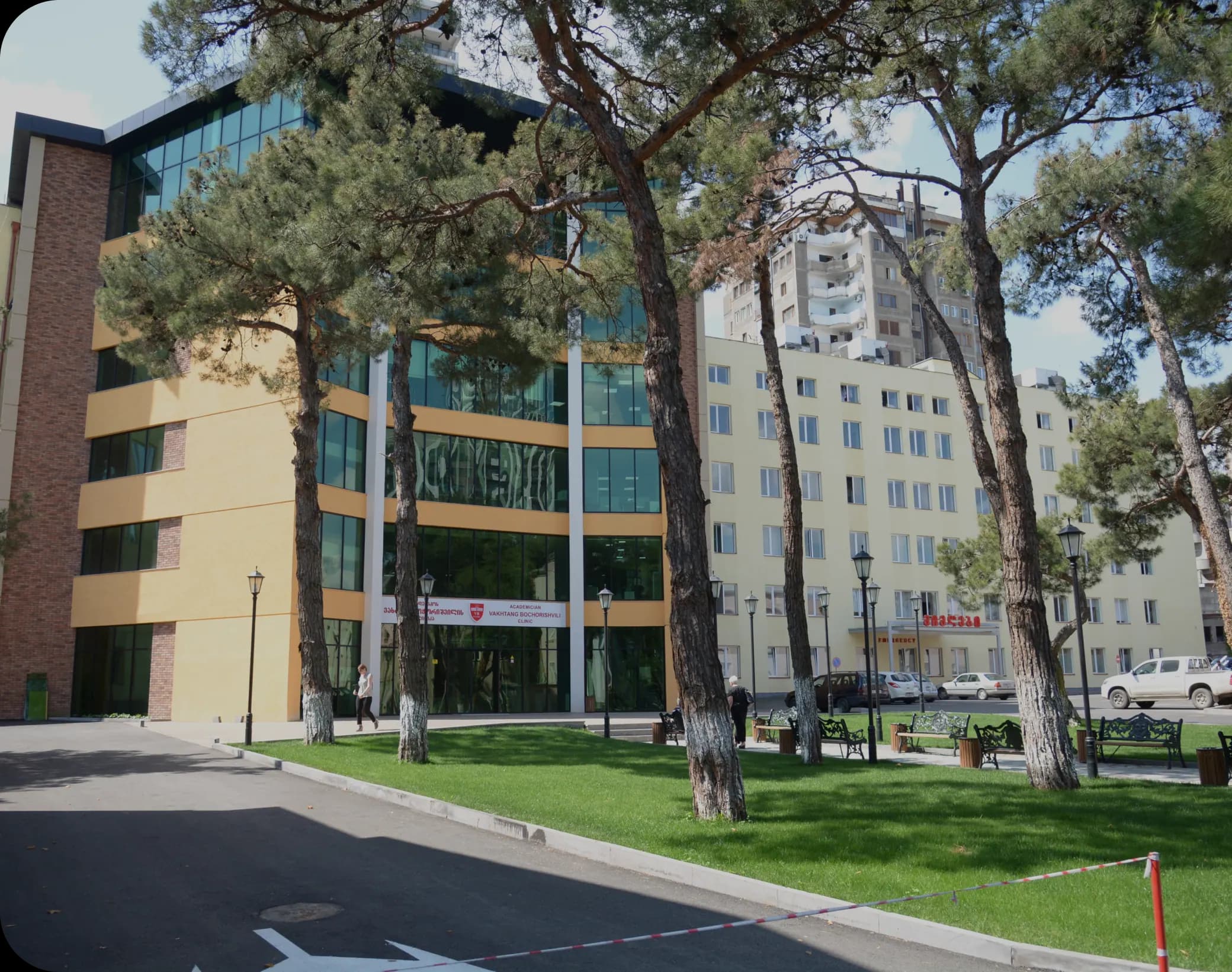
Magnetic resonance imaging (MRI) of the spine at Bochorishvili Clinic with the Siemens MAGNETOM Vida 3T machine. Learn more about the study.
Back pain, pain radiating to the extremities, numbness, or limited movement are often associated with various spinal diseases. One of the most informative studies for accurate diagnosis of such conditions is magnetic resonance imaging (MRI) of the spine.
MRI allows the doctor to assess the vertebrae, intervertebral discs, spinal cord, nerve roots, and surrounding soft tissues in detail. The study does not use ionizing radiation and, through high-quality imaging, facilitates the early detection of pathological changes.
When is a spine MRI prescribed?
A spine MRI is prescribed when it is necessary to determine the cause of back pain or neurological symptoms.
A spine MRI may be ordered if the patient has:
- Prolonged or severe pain in the neck, chest, or lower back;
- Spreading pain into the arm or leg;
- Numbness of the limbs;
- Tingling sensation;
- Muscle weakness;
- Restriction of movement;
- Trauma;
- Suspicion of a herniated disc;
- Suspicion of spinal canal narrowing;
- Urination or defecation disorders accompanied by neurological symptoms.
What diseases can it detect?
MRI of the spine allows the detection of degenerative, inflammatory, traumatic, and tumor diseases. The study is most often used to diagnose the following pathologies:
- Disc herniation (hernia) - damage to a disc when it puts pressure on nerve roots or the spinal cord.
- Disc protrusion - a bulging of the disc, which is often considered the first stage of a hernia.
- Spinal stenosis - narrowing of the spinal canal, which causes compression of nerve structures.
- Degenerative changes - age- or overload-related changes in the vertebrae and intervertebral discs.
- Spondylosis - a degenerative disease of the spine that often causes bone spurs and restricted movement.
- Spondylolisthesis - a condition where one vertebra is displaced forward or backward relative to the other.
- Spinal cord injuries - traumatic, degenerative, or other changes that damage the spinal cord.
- Tumors and metastases - both primary and tumor processes that spread from other organs.
- Infectious processes - infections of the spine, intervertebral discs, or surrounding tissues.
- Inflammatory diseases - including chronic inflammatory changes that affect various structures of the spine.
- Traumatic injuries - fractures, ligament damage, soft tissue trauma and other injuries.
How is the research conducted?
Before the examination begins, the patient removes all metal objects and lies on the moving table of the CT scanner. Depending on which part of the spine is being examined, the patient is placed in the appropriate position to ensure the most accurate image.
During the examination, the table is slowly moved into the CT scanner, where a special magnetic field and radio waves are used to produce detailed images of the spine. It is important that the patient lie as still as possible during the procedure, as movement can reduce the quality of the image.
The device emits a characteristic sound during the examination. Therefore, if necessary, the patient will be given headphones or earplugs.
In some cases, if a more detailed assessment of blood vessels, tumors, or inflammatory processes is needed, the study is performed using a contrast agent, which is injected into a vein by medical personnel before or during the study.
The duration of a spine MRI depends on which part of the spine is being examined, but the procedure usually lasts 15-45 minutes.
How to prepare for an MRI scan?
No special preparation is usually required before the test. However, to ensure the test is conducted safely, it is important to inform your doctor in advance about your health condition and any medical devices you have.
Be sure to tell your doctor if:
- Do you have a pacemaker or other electronic implant?
- You have a metal implant, prosthesis or metal fragment in your body;
- Are you pregnant or suspect you are pregnant?
- You have had an allergic reaction to the contrast material used during an MRI in the past.
In the case of a contrast study, the doctor may also make additional recommendations.
Siemens MAGNETOM Vida 3T - the latest generation tomograph at Bochorishvili Clinic
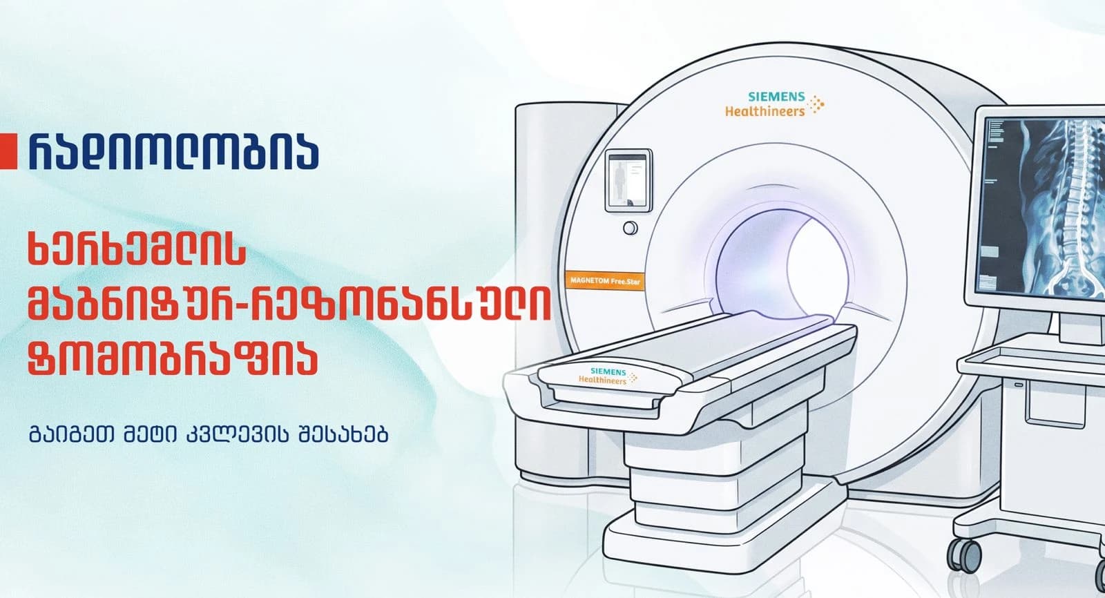
The clinic performs spine MRI using the Siemens MAGNETOM Vida 3T machine - a new generation 3-Tesla magnetic resonance imaging scanner.
The main advantages of the device are:
- 3-Tesla magnetic field, which provides high-resolution and detailed images;
- BioMatrix technology automatically adapts to the patient's individual anatomical features and helps to obtain more accurate results;
- Fast scanning modes that reduce examination time;
- Artificial intelligence-based technologies improve image quality and optimize the research process;
- 70 cm wide tunnel for maximum comfort during the examination, including for patients with claustrophobia;
- Integrated video screen, which allows the patient to watch the desired video during the examination;
- An orthopedic mattress reduces discomfort and ensures a comfortable position during the examination.
These technological capabilities are especially important when it comes to accurately assessing small changes in the intervertebral discs, spinal cord, nerve roots, and other spinal structures, which is crucial for correct diagnosis and treatment planning.
What is the difference between a spine MRI and CT?
MRI (magnetic resonance imaging) and CT (computed tomography) are both diagnostic tests, but they provide different information.
MRI shows the intervertebral discs, spinal cord, nerve roots, ligaments, and other soft tissues in the most detailed way. Therefore, it is often used to detect herniated discs, protrusions, nerve compression, and other similar pathologies. MRI uses a strong magnetic field and radio waves to produce detailed images.
CT provides better evaluation of bony structures. It is particularly informative in evaluating fractures, bone lesions, and some structural changes. Computed tomography uses X-rays and the examination process is shorter than MRI.
Which test is needed depends on your symptoms and the likely diagnosis. Your doctor will select the appropriate test based on your clinical assessment.
Conclusion
Magnetic resonance imaging (MRI) shows what is happening in the vertebrae, intervertebral discs, spinal cord, and nerve structures. The information obtained from this study helps the doctor correctly assess the patient's condition and plan further steps.
To book the examination, contact us:
032 2 312 112 | 577 22 21 12
📍 Al. Kazbegi 16, Tbilisi
Frequently Asked Questions