 ქართული
 English
 Русский
Blog
Dissolving kidney stones
Dissolving kidney stones
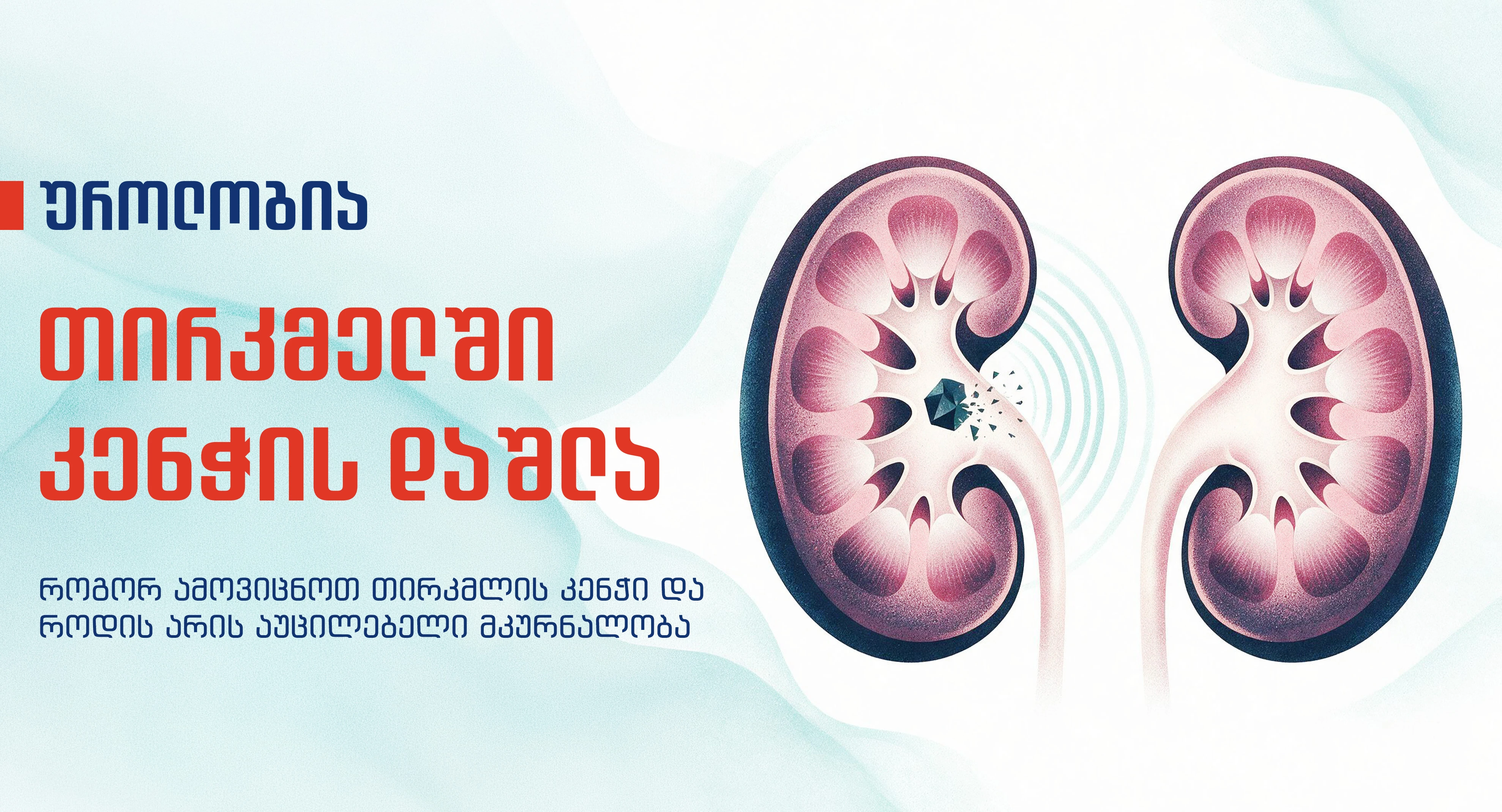
Kidney stones (nephrolithiasis) are one of the most common urological problems that significantly affect human health. The disease can manifest with various symptoms and develop at any age.
Kidney stones can cause severe pain, urinary tract infections, and in some cases, kidney failure. That's why early diagnosis and appropriate treatment are especially important.
What is a kidney stone?
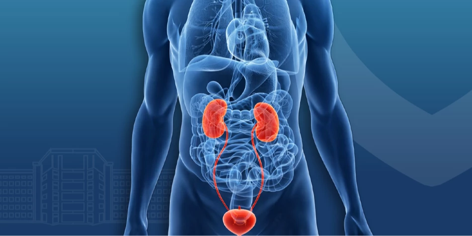
The kidneys filter the blood and remove excess fluid and substances as urine. When the concentration of certain minerals (calcium, oxalate, or uric acid) in the urine increases, they begin to form crystals. Over time, these crystals stick together and form stones.
Small stones can sometimes pass naturally from the body and do not require any special intervention. However, larger stones can cause severe pain, infection, and damage to kidney function.
Kidney stones can develop:
- In the kidney;
- In the urethra;
- In the bladder.
Their size ranges from a few millimeters to several centimeters. The larger the stone, the greater the risk of developing complications.
What causes kidney stones to form?
There is usually no single cause for the development of kidney stones. In most cases, the disease is the result of a combination of factors.
The following factors increase the risk of developing kidney stones:
Insufficient fluid intake
Dehydration is one of the biggest risk factors for developing kidney stones. When a person doesn't drink enough water, their urine becomes more concentrated, which encourages the formation of crystals.
Eating habits
Certain dietary habits may increase the risk of developing stones, including:
- Excessive salt intake;
- Consuming large amounts of animal protein;
- Excessive intake of products rich in oxalates (spinach, beets, almonds, chocolate, etc.).
Hereditary factors
If someone in your family has had kidney stones, you are more likely to develop the disease. Genetic factors may determine your body's predisposition to developing certain types of stones.
Metabolic disorders
In some cases, stone formation is associated with metabolic diseases, such as:
- with calcium metabolism disorders;
- With increased uric acid levels;
- With certain diseases of the parathyroid gland.
Urinary tract infections
Recurrent urinary tract infections may contribute to the formation of certain types of kidney stones. Infection changes the acidity and composition of urine, which favors the formation of stones.
What are the symptoms of kidney stones?
Small stones are often asymptomatic. Symptoms usually develop when the stone moves into the ureter and blocks the flow of urine.
The most common signs of the disease are:
- Severe, sudden pain in the lower back or side;
- Pain spreading from the lower abdomen and towards the groin;
- Blood in the urine;
- Frequent or painful urination;
- Nausea and vomiting;
- Discomfort and restlessness.
When a stone moves into the ureter, the risk of developing renal colic increases , which is characterized by severe, stabbing pain in the lower back or side.
If these symptoms are accompanied by a high fever, chills, or difficulty urinating, it is necessary to see a doctor immediately, as this indicates an infection or other serious complications.
How is a kidney stone diagnosed?
To make an accurate diagnosis, a urologist evaluates the patient's complaints, medical history, and, if necessary, prescribes additional tests. In addition to determining the presence of a stone, the goal of diagnostics is to assess its size, location, number, and possible complications, which helps the doctor choose the right treatment tactics.
The following tests are used to diagnose kidney stones:
- Urologist consultation;
- General urine analysis;
- Blood laboratory tests;
- Ultrasound examination of the kidneys and urinary system;
- Computed tomography (CT).
Computed tomography (CT) is one of the most accurate methods for detecting kidney stones. The study allows for the assessment of the exact location, size, number of stones, and their possible impact on the urinary tract. The information obtained is important for selecting the optimal treatment method.
Kidney stone dissolution and treatment
The treatment for kidney stones is determined individually and depends on several important factors. The doctor takes into account the size, location, composition of the stone, the condition of the urinary tract, the patient's symptoms, and their general health. Small stones can sometimes pass naturally from the body, but larger or more complicated cases require special intervention.
The main methods of treating kidney stones are:
Conservative treatment
If the kidney or ureteral stone is small and has a high probability of passing naturally, conservative treatment is preferred. This approach is used in cases where the stone does not obstruct urination, does not cause kidney function impairment, infection, or other complications.
The main goal of conservative treatment is to control pain and other symptoms, promote natural passage of the stone, and prevent complications.
During the treatment period, the patient is under the supervision of a doctor, and if necessary, repeated examinations are performed to assess the location and movement of the stone.
Conservative treatment for gallstones includes:
- Drink plenty of fluids - Hydration increases urine volume, which in some cases helps to naturally pass small stones.
- Drug therapy to aid in stone passage - in certain cases, medications are prescribed that reduce spasm of the muscular wall of the ureter, help it relax, and facilitate the natural passage of the stone.
- Periodic monitoring - Regular assessment of the patient's condition is important during the treatment process. The doctor will most likely order a repeat ultrasound, CT scan, or other diagnostic examination.
If, despite conservative treatment, the stone is not expelled from the body, the pain increases, an infection develops, urinary flow is obstructed, or kidney function is impaired, other treatment methods may be necessary, such as remote or laser lithotripsy and surgery.
Extracorporeal shock wave lithotripsy (ESWL)
Extracorporeal Shock Wave Lithotripsy (ESWL) is a modern, non-surgical method in which special shock waves impact the stone from outside the body, breaking it into small fragments, which are then naturally excreted from the body with urine.
Remote lithotripsy gives the best results in the case of small and medium-sized stones - up to 2 cm. Especially when they are localized in the kidney or upper part of the ureter. The doctor assesses the feasibility of the procedure taking into account the size, location, density of the stone and the patient's general health.
The main advantages of remote lithotripsy are:
- Does not require a surgical incision - the procedure is performed without damaging the skin, which reduces the risks associated with surgery.
- The procedure lasts an average of 30–60 minutes - in most cases it can be performed in a shorter time.
- Rapid recovery period - most patients return to daily activities in a short time.
- Short-term medical supervision - In most cases, hospitalization is not necessary or is very short-term.
- Low risk of complications - The likelihood of complications is much lower compared to open surgeries.
The stone, broken into small fragments by remote lithotripsy, is eventually excreted in the urine. This process may take several days, several weeks, or, in some cases, longer.
Laser lithotripsy
If the stone is small but cannot be passed on its own, causes severe pain, obstructs urine flow, or is located in a location that is less favorable for remote lithotripsy, laser lithotripsy is often an effective solution for dissolving the stone.
Laser lithotripsy is a modern, minimally invasive endoscopic method. During the procedure, the doctor reaches the stone through the urinary tract with a special thin endoscope and uses laser energy to break it into small fragments. The broken fragments are then removed by the doctor with the help of special instruments.
The main advantages of laser lithotripsy are:
- Direct impact on the stone - laser energy directly affects the stone, allowing the doctor to effectively break it up.
- Used for various types of stones - the method is used to treat stones of different sizes and densities.
- It is effective in cases of difficult localization - when the stone is in a part of the kidney or ureter where remote lithotripsy would be less effective.
- It is a minimally invasive procedure - the procedure is performed through the urinary tract and usually does not require a surgical incision.
- Rapid recovery period - most patients return to daily activities in a short time.
In some cases, after the procedure, the doctor temporarily places a ureteral stent (Double J stent) to prevent the free flow of urine and to help pass any remaining small stone fragments. The need for the stent and how long it will be left in place are determined individually.
In the case of very large and high-density stones, percutaneous nephrolithotomy (PCNL) is the most advantageous.
Percutaneous nephrolithotomy (PCNL)
Percutaneous Nephrolithotomy (PCNL) is a modern, minimally invasive surgical method used in cases where remote lithotripsy or other less invasive methods are not effective.
During the procedure, a small incision is made in the lumbar region, through which special instruments are inserted directly into the kidney. The doctor dislodges the stone and removes its fragments with special instruments, which in most cases allows the kidney to be completely freed.
This method is particularly effective for stones larger than 2 cm , coral-like stones, multiple stones, or stones that cannot be dissolved or completely removed by other methods due to their difficult location.
The main advantages of percutaneous nephrolithotomy are:
- High efficiency in case of large stones - in most cases, it is possible to completely dissolve and remove the stone within a single procedure.
- It is a minimally invasive method - the operation is performed through a small incision, which reduces tissue damage.
- Precise visual control - During the procedure, the surgeon directly sees the stone, which allows for its effective disintegration and removal.
After the procedure, the patient remains under the supervision of doctors for several days. The recovery period depends on the individual condition of the patient.
Following your doctor's recommendations and following up with your doctor is important for a full recovery and preventing the recurrence of stones.
How to reduce the risk of kidney stones?
It is not always possible to completely prevent all cases of kidney stones, as their development is influenced by lifestyle, genetic, and metabolic factors. However, by following certain recommendations, it is possible to significantly reduce the likelihood of stone formation and prevent recurrence.
For prevention it is important to:
- Drink enough water - drinking adequate amounts of fluids throughout the day helps dilute urine and reduces the risk of accumulation of substances that contribute to stone formation;
- Reducing salt intake - excess salt contributes to an increase in the amount of calcium in the urine, which is one of the factors in the formation of stones;
- Balanced diet - a varied diet is recommended, including vegetables, fruits, sufficient protein and calcium according to the doctor's recommendations;
- Maintaining a healthy body weight - being overweight and having metabolic disorders may increase the risk of developing kidney stones;
- Regular physical activity - movement has a positive effect on metabolism, which reduces the likelihood of stone formation.
If kidney stones recur, your doctor will order additional tests, including urine and blood tests, to assess factors that contribute to stone formation and develop a prevention plan.
Kidney stone removal at Bochorishvili Clinic
When treating kidney stones, it is important to choose the right method that will be effective considering the specific patient's condition, size, and location of the stone. At Bochorishvili Clinic, kidney stones are treated using modern approaches.
In addition to conservative treatment, the following methods of dissolving kidney stones are available at the clinic:
- Extracorporeal shock wave lithotripsy (ESWL) is a non-invasive method in which stones are broken up using shock waves, without surgical intervention.
Bochorishvili Clinic uses the latest generation remote lithotripsy device - STORZ MEDICAL MODULITH SLX-F2 for this procedure . The device allows the doctor to Clear and detailed images that ensure maximum accuracy during the procedure. - Laser lithotripsy - an endoscopic method in which the stone is broken up with a laser. It is effective in cases of stones of various sizes and locations.
- Percutaneous nephrolithotomy (PCNL) - Used in cases where the size, location, or other characteristics of the stone require minimally invasive intervention.
The selection of treatment tactics is preceded by an accurate diagnosis, which allows for the assessment of the characteristics of the stone and the determination of the most effective treatment method for the patient.
Bochorishvili Clinic performs extracorporeal shock wave lithotripsy (ESWL), laser lithotripsy, and percutaneous nephrolithotomy (PCNL):
- Head of the Urology Department Nikoloz Gogoladze
- Urologist Bakar Machitidze
Conclusion
Kidney stones can be a painful and distressing condition, but with modern medicine, they can be effectively managed and treated. Successful management of the condition involves not only removing the stone, but also assessing the cause of its formation and reducing the risk of recurrence.
Modern diagnostic methods allow the doctor to accurately determine the size, location, and composition of the stone and plan treatment based on these factors.
Ignoring the symptoms of kidney stones can lead to complications. If you are experiencing severe pain in the lower back, blood in the urine, or other suspicious signs, it is important to consult a urologist in a timely manner. Early diagnosis allows for a timely assessment of the condition, the correct choice of treatment tactics, and the prevention of possible complications.